Our website is made possible by displaying online advertisements to our visitors.
Please consider supporting us by disabling your ad blocker.
Third ventricle
This article needs additional citations for verification. (November 2007) |
| Third ventricle | |
|---|---|
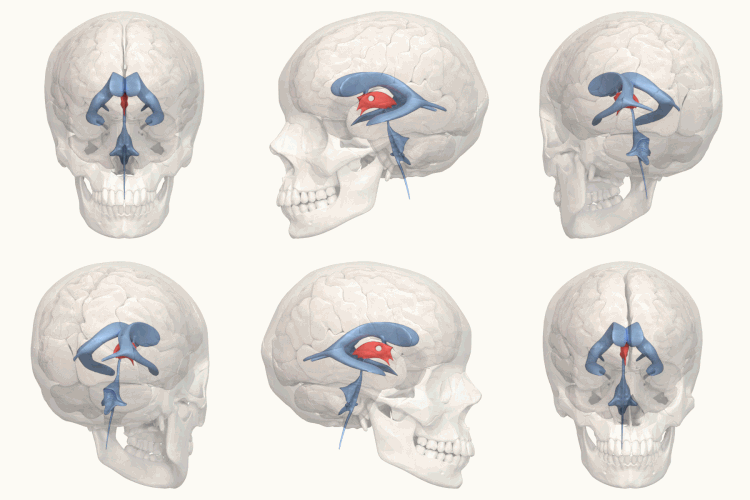
 Third ventricle shown in red | |
 Blue – lateral ventricles Cyan – interventricular foramina (Monro) Yellow – third ventricle Red – cerebral aqueduct (Sylvius) Purple – fourth ventricle Green – continuous with the central canal (apertures to subarachnoid space are not visible) | |
| Details | |
| Identifiers | |
| Latin | ventriculus tertius cerebri |
| MeSH | D020542 |
| NeuroNames | 446 |
| NeuroLex ID | birnlex_714 |
| TA98 | A14.1.08.410 |
| TA2 | 5769 |
| FMA | 78454 |
| Anatomical terms of neuroanatomy | |
The third ventricle is one of the four connected cerebral ventricles of the ventricular system within the mammalian brain. It is a slit-like cavity formed in the diencephalon between the two thalami, in the midline between the right and left lateral ventricles, and is filled with cerebrospinal fluid (CSF).[1]
Running through the third ventricle is the interthalamic adhesion, which contains thalamic neurons and fibers that may connect the two thalami.
- ^ Singh, Vishram (2014). Textbook of Anatomy Head, Neck, and Brain; Volume III (2nd ed.). Elsevier. pp. 386–387. ISBN 9788131237274.
Previous Page Next Page


